Tissue Under Pressure: The Cellular Mechanics of What Prolonged Sitting Does to Your Spine, Hips, and Connective Tissue
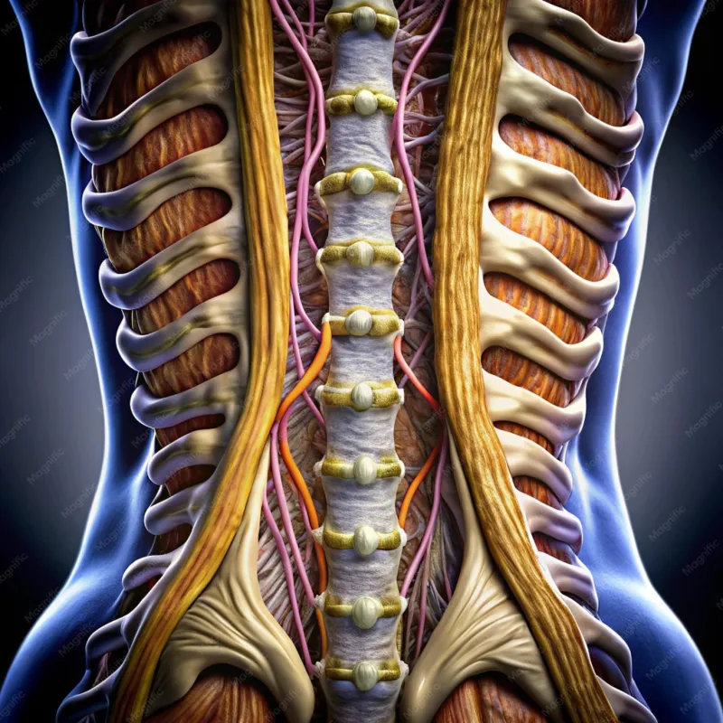
Photo: human spine anatomy cross section posture desk worker office, via img.freepik.com
The average American office worker spends somewhere between nine and twelve hours per day in a seated position. Public health messaging has spent years repeating the phrase "sitting is the new smoking," yet that comparison, however attention-grabbing, tells us very little about the underlying biology. What is actually happening inside the body when a person remains seated for sustained periods? The answer involves collagen remodeling, fluid dynamics in cartilage, adaptive shortening of contractile proteins, and a cascade of biomechanical compensations that the nervous system encodes as its new definition of "normal."
This is not a story about laziness or poor discipline. It is a story about tissue responding rationally—and often permanently—to the mechanical environment it is placed in, day after day.
The Disc as a Hydraulic System
The intervertebral discs that cushion each vertebral segment of the spine are not passive spacers. They function as dynamic, fluid-filled structures whose integrity depends on cyclical loading and unloading. The nucleus pulposus—the gel-like interior of each disc—maintains its height and shock-absorbing capacity through a process called imbibition, in which movement draws water and nutrients into the disc from surrounding tissue.
When a person sits for extended periods, particularly with the lumbar spine in a flexed or slouched position, the compressive load on the anterior portion of the disc becomes chronically elevated. This sustained compression drives fluid out of the nucleus pulposus without the corresponding decompression phase that movement provides. Over time, the disc loses hydration, reduces in height, and becomes stiffer and less capable of distributing mechanical load evenly across the vertebral endplate.
Research using MRI imaging has demonstrated measurable disc height loss after as few as four hours of continuous sitting. While some of this fluid loss is recovered with recumbency and movement, years of repeated compression without adequate recovery create cumulative changes in disc matrix composition—specifically a reduction in proteoglycan content, the molecules responsible for water retention within the disc. The result is a structure that is biochemically older than the chronological age of the person it belongs to.
Fascia Does Not Forget
Deep to the skin and surrounding every muscle, bone, and organ lies the fascial network—a continuous web of collagen-rich connective tissue that transmits mechanical force across the body. Fascia is not inert. It contains fibroblasts, the cells responsible for synthesizing and degrading collagen, and these cells are exquisitely sensitive to mechanical input.
When sustained pressure or immobility is applied to a fascial region, fibroblasts respond by increasing collagen deposition in the direction of habitual loading. This process, sometimes referred to as mechanotransduction-driven remodeling, is the body's attempt to reinforce tissue under repeated stress. In a healthy, mobile body, this remodeling is balanced and adaptive. In a chronically sedentary one, it produces fascial thickening, reduced tissue glide between adjacent layers, and the formation of cross-links between collagen fibers that restrict range of motion.
The thoracolumbar fascia—a particularly dense sheet spanning the lower back—is among the most affected regions in habitual sitters. As this structure thickens and loses its capacity for smooth mechanical transmission, it begins to restrict hip extension and lateral movement of the lumbar spine. Individuals often perceive this as tightness or stiffness, though the actual mechanism is structural rather than purely muscular.
Adaptive Shortening and the Psoas Problem
Muscle tissue, like fascia, adapts to the position in which it spends the most time. The iliopsoas—a deep hip flexor that originates on the lumbar vertebrae and inserts on the femur—is held in a shortened position throughout every hour of seated work. Over weeks and months, the muscle responds by reducing the number of sarcomeres in series within each myofibril. This is known as adaptive shortening, and it represents a genuine architectural change in the contractile unit of the muscle.
A psoas that has undergone adaptive shortening does not simply feel tight. It exerts a persistent anterior pull on the lumbar vertebrae and the femur, even at rest. When the person stands, the muscle cannot achieve full elongation, and the pelvis compensates by tilting anteriorly—a posture commonly described as anterior pelvic tilt. This tilt increases the lumbar lordotic curve, compresses the posterior elements of the lumbar spine, and alters the mechanical demands placed on the gluteal musculature.
The gluteus maximus, the body's primary hip extensor and one of its most powerful stabilizers, is simultaneously inhibited through a process called reciprocal inhibition. When the hip flexors are chronically shortened and tonically active, the nervous system reduces motor drive to the opposing gluteal muscles. This phenomenon—sometimes called gluteal amnesia in clinical contexts—means that the largest muscle group in the lower body gradually loses its ability to generate force efficiently, shifting mechanical burden to the hamstrings, lumbar erectors, and even the knee joint.
The Cervical Spine and the Weight of a Forward Head
The effects of prolonged sitting are not confined to the lumbar region and hips. The position most people adopt when working at a desk or looking at a screen involves a degree of forward head posture—the skull translating anteriorly relative to the cervical spine. For every inch the head moves forward from its neutral position over the shoulders, the effective load on the cervical spine increases by approximately ten pounds, according to biomechanical modeling research.
This added load is absorbed by the posterior cervical musculature—the suboccipitals, semispinalis, and upper trapezius—which must contract continuously to prevent the head from falling further forward. Sustained isometric contraction in these muscles leads to localized ischemia, accumulation of metabolic byproducts, and eventually the formation of myofascial trigger points. The cervical vertebrae themselves experience altered loading patterns, which over time can accelerate degenerative changes in the facet joints and disc spaces of the upper spine.
Why Compensation Patterns Persist After Movement Resumes
One of the most clinically significant aspects of sedentary-induced tissue remodeling is that it does not immediately reverse when a person begins exercising. The nervous system encodes habitual posture as a reference position—a process mediated through the gamma motor neuron system, which sets baseline muscle tone by adjusting the sensitivity of muscle spindles. When the brain has recalibrated its positional reference to accommodate anterior pelvic tilt and forward head posture, it will actively maintain those positions even during movement.
This is why an individual who begins a new exercise program after years of desk work often finds that their squat mechanics are compromised, their running gait is asymmetrical, or their shoulder mobility is limited despite weeks of stretching. The problem is not simply muscular tightness—it is a neurological template that must be systematically reconditioned through specific corrective input.
Rethinking the Sedentary Problem
Addressing the physiological consequences of chronic sitting requires more than occasional walks or a standing desk purchased out of guilt. It demands an understanding of which tissues are being remodeled, in which direction, and over what time course. Targeted fascial mobilization, sarcomere lengthening protocols for the hip flexors, and deliberate neuromuscular re-education for the gluteal complex represent categories of intervention grounded in the actual mechanisms described above.
The body is not being damaged by sitting in any single session. It is being gradually reshaped by the cumulative mechanical environment it inhabits. That is both the sobering reality and the hopeful one—because environments, unlike genetics, can be deliberately altered. Understanding the tissue-level story is the necessary first step.