From Damage to Recovery: A Cellular Timeline of the Inflammatory Response
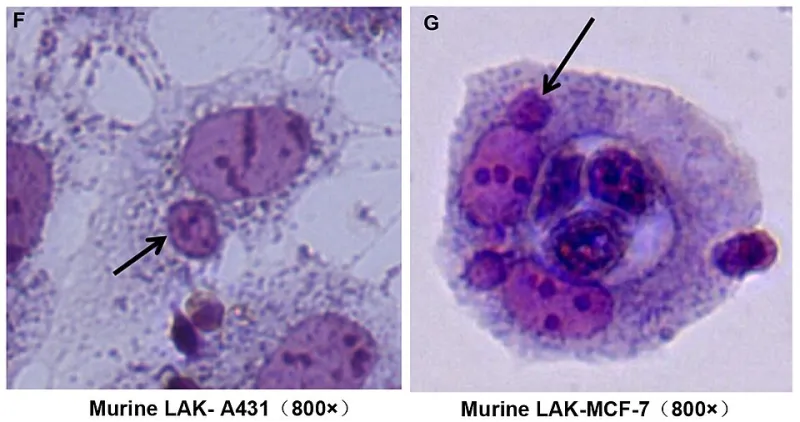
Photo: Chen Y-h, Wang S, He M-f, Wang Y, Zhao H, Zhu H-y, et al., CC BY 4.0, via Wikimedia Commons
Open any wellness publication or scroll through fitness content on social media, and you will encounter inflammation treated as an unambiguous enemy—something to be suppressed with ice baths, anti-inflammatory supplements, and targeted pharmaceuticals at every opportunity. This framing, while culturally pervasive, fundamentally misrepresents what inflammation actually is: a tightly regulated, sequentially organized biological program without which wounds would never close and infections would go unchecked. To understand recovery, one must first understand the cascade.
The Trigger: Injury at the Tissue Level
Inflammation begins not with redness or swelling, but with cellular damage. Whether the instigating event is a sprained ankle on a basketball court, a muscle tear from a heavy squat, or a laceration, the immediate consequence is disruption of cell membranes and the release of intracellular contents into the surrounding tissue. Damaged cells release a collection of signaling molecules—including prostaglandins, histamine, and bradykinin—that serve as the body's first alarm system.
Within seconds to minutes, these chemical signals trigger local blood vessels to dilate, increasing blood flow to the affected area. Simultaneously, the vessels become more permeable, allowing plasma proteins and immune cells to leak into the tissue space. This is the biological origin of the four cardinal signs of acute inflammation that physicians have recognized since antiquity: redness (rubor), heat (calor), swelling (tumor), and pain (dolor). Each of these signs reflects an adaptive mechanism, not a malfunction.
The First Responders: Neutrophils Arrive
Within the first hour following injury, neutrophils—the most abundant white blood cells in human circulation—begin arriving at the site in significant numbers. These cells are the immune system's rapid-response infantry. They are drawn to the injury by chemotactic signals, molecular breadcrumbs that guide them through vessel walls and into the damaged tissue via a process called diapedesis.
Neutrophils serve two immediate functions. First, they phagocytize—engulf and destroy—pathogens, debris, and damaged cell fragments. Second, they release additional inflammatory mediators that amplify the local response and recruit subsequent immune players. This amplification is intentional: the body is escalating its response proportionally to the scale of the damage detected.
This phase typically peaks between 24 and 48 hours post-injury. It is during this window that swelling and pain are most pronounced, and it is also the phase most commonly targeted by interventions such as ice application and nonsteroidal anti-inflammatory drugs (NSAIDs).
The Ice vs. Heat Question, Answered by Biology
The debate over ice versus heat in injury management is most productively resolved by anchoring the decision to this cellular timeline. During the acute inflammatory phase—roughly the first 48 to 72 hours—ice (cryotherapy) can serve a legitimate purpose. By constricting blood vessels and reducing nerve conduction velocity, cold application decreases swelling accumulation and provides meaningful analgesia. It does not stop inflammation, but it modulates the magnitude of the vascular response.
Heat, by contrast, promotes vasodilation and increases metabolic activity in tissues. Applied during the acute phase, heat can exacerbate swelling and accelerate cellular processes that are already operating at maximum capacity. It becomes appropriate—indeed, beneficial—during the subacute and chronic phases of healing, when increased blood flow supports tissue remodeling and reduces muscular stiffness.
The error that many athletes and recreational exercisers make is applying ice reflexively for days or weeks after an injury, inadvertently blunting the later phases of healing that depend on robust circulation and cellular activity.
The Macrophage Transition: Inflammation Becomes Repair
Approximately 48 to 96 hours after injury, the cellular composition of the inflammatory site begins to shift. Neutrophils, having completed their initial work, undergo programmed cell death. Macrophages—larger, longer-lived immune cells—move to the forefront and assume a fundamentally different role.
Macrophages are phenotypically plastic, meaning they can adopt different functional profiles depending on the local chemical environment. In the early post-injury period, they express what is called an M1 phenotype: pro-inflammatory, aggressively phagocytic, and oriented toward pathogen clearance. As the acute phase resolves, they transition toward an M2 phenotype, which is characterized by the secretion of growth factors, including transforming growth factor-beta (TGF-β) and vascular endothelial growth factor (VEGF), that stimulate tissue repair and angiogenesis—the formation of new blood vessels.
This macrophage transition is among the most critical junctures in the entire healing cascade. Research conducted over the past two decades has demonstrated that disrupting this transition—through excessive use of NSAIDs, for example—can impair tendon and muscle healing by interfering with the growth factor signaling that drives tissue remodeling.
Proliferation: Building the Scaffold
From roughly day three through week three, depending on injury severity, the body enters the proliferative phase. Fibroblasts—cells specialized in producing structural proteins—migrate into the wound site and begin synthesizing collagen. This collagen initially forms a disorganized matrix that provides mechanical strength sufficient to hold tissue together, but which lacks the precise architectural alignment of uninjured tissue.
Simultaneously, new capillaries infiltrate the healing tissue, and epithelial cells proliferate to resurface any disrupted skin or mucosal layers. The wound contracts as myofibroblasts—a hybrid cell type with properties of both fibroblasts and smooth muscle cells—pull the wound edges together.
This phase is when rehabilitation exercise, if appropriately dosed, plays a profoundly constructive role. Controlled mechanical loading during the proliferative phase sends tensional signals to fibroblasts that guide collagen fiber alignment along the axes of functional stress. Immobilization, by contrast, results in randomly oriented collagen deposition that is weaker and more prone to re-injury.
Resolution: The Underappreciated Final Chapter
The resolution phase—sometimes lasting weeks to months—involves the remodeling of immature scar tissue into more organized, load-bearing connective tissue. Collagen cross-linking increases, excess cells undergo apoptosis, and the tissue gradually recovers tensile strength. Specialized lipid mediators called resolvins and protectins, derived from omega-3 fatty acids, actively facilitate this resolution by suppressing residual inflammatory signaling.
When this phase is disrupted—by chronic re-injury, persistent infection, or metabolic factors such as poor glycemic control—inflammation fails to resolve and transitions into a chronic state. Chronic inflammation is characterized by persistent low-grade immune activation, ongoing tissue degradation, and impaired repair. This is the form of inflammation that is legitimately associated with long-term health consequences, and it is fundamentally different in character from the acute, purposeful response described above.
What This Means for Recovery Practice
Understanding inflammation as a timeline rather than a static condition transforms how one approaches recovery. Suppressing the acute phase indiscriminately delays healing. Failing to support the proliferative phase with appropriate movement and nutrition prolongs it unnecessarily. Ignoring the resolution phase entirely—as many athletes do once pain subsides—leaves tissue incompletely remodeled and vulnerable to recurrence.
Nutrition plays a supporting role throughout. Adequate protein intake provides the amino acid substrates for collagen synthesis. Vitamin C is an essential cofactor in collagen cross-linking. Zinc supports immune cell function during the early phases. And omega-3 fatty acids, through their conversion to resolvins and protectins, actively accelerate resolution.
The body's inflammatory cascade is not an adversary to be defeated. It is a precisely timed biological program that, when supported rather than suppressed, represents the most sophisticated healing technology available to the human organism.